Gastrointestinal Cancers


Gastric cancer (Stomach cancer)

Pathogenesis & Risk Factors of Gastric Adenocarcinoma
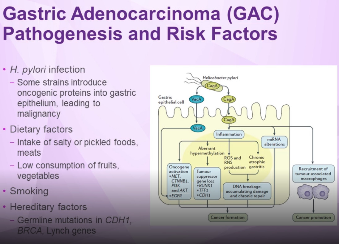
Pathogenesis & Risk Factors of Gastric Adenocarcinoma

Some strains of H. pylori can introduce oncogenic proteins into the gastric epithelium; cytotoxin-associated gene A (CagA) can induce activation of oncogenes (MET, CTNNB1, PI3K, and AKT), aberrant hypermethylation resulting in loss of tumor suppressor genes (RUNX1, TFF1, and CDH1), and increased levels of reactive oxygen species (ROS), and reactive nitrogen species (RNS).1 Vacuolating cytotoxin A (VacA) activates eGFR, which also promotes cancer formation.

Molecular Subtypes of Gastric Cancer
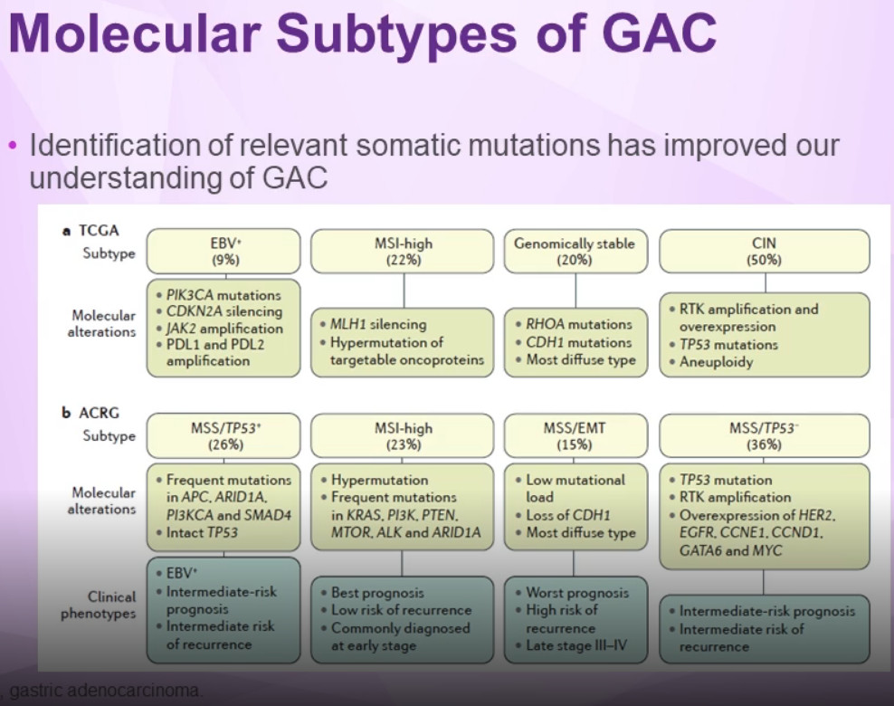
Molecular Subtypes of Gastric Cancer


Actionable Biomarkers of Gastric Adenocarcinoma
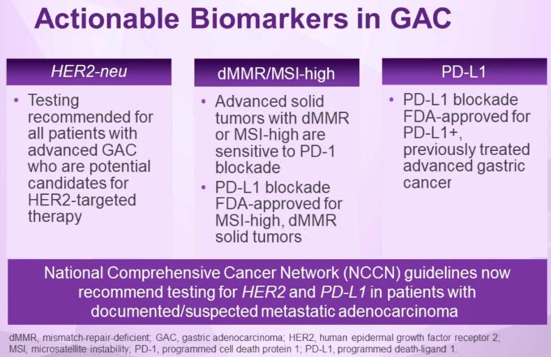
Actionable Biomarkers of Gastric Adenocarcinoma

NCCN Guildliness for Treatment of Gastric Adenocarcinoma
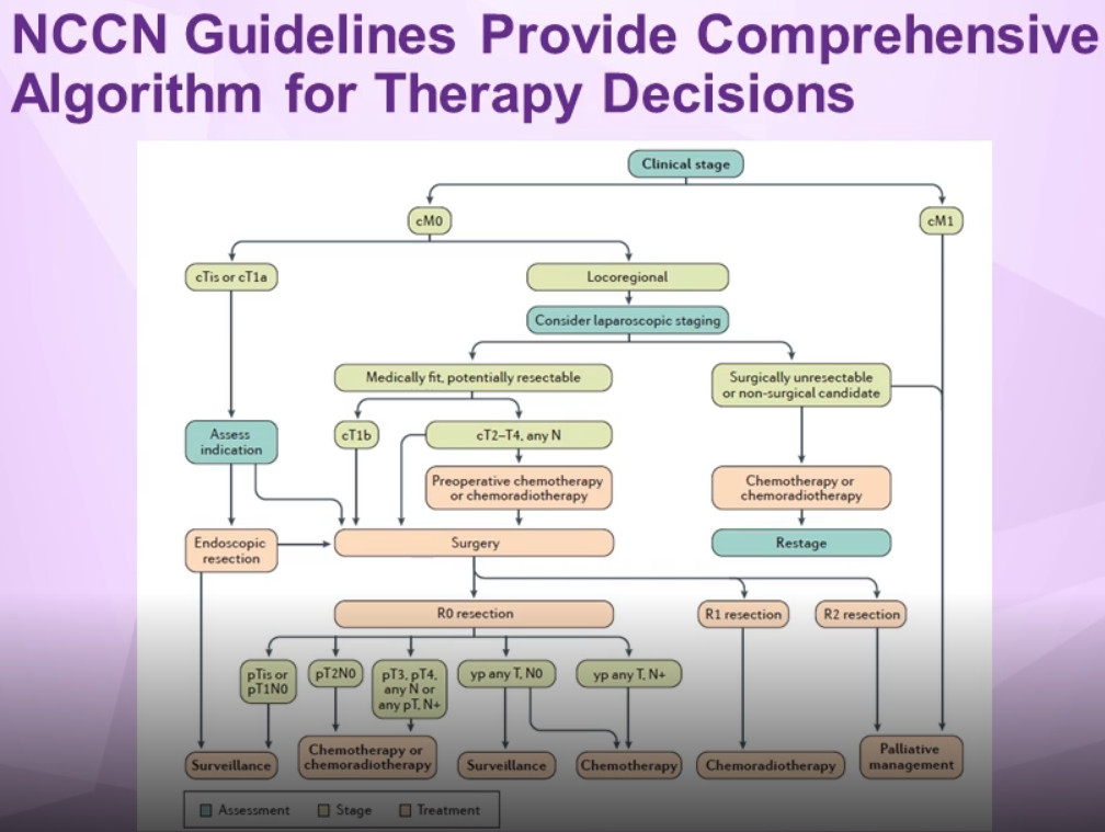
NCCN Guildliness for Treatment of Gastric Adenocarcinoma


Treatment of gastric cancer

Stomach cancer may be treated with surgery, radiation therapy, chemotherapy, targeted therapy, or immunotherapy... Often, a combination of these treatments is used. It can be difficult to cure stomach cancer because it is often not found until it is at an advanced stage.

Treatment options and recommendations depend on several factors, including the type and stage of cancer, possible side effects, and the patient’s preferences and overall health. Your care plan may also include treatment for symptoms and side effects, an important part of cancer care.

Surgery
The type of surgery used depends on the stage of the cancer (see Stages).
For a very early stage (T1a) cancer, some doctors may recommend a non-surgical treatment called endoscopic mucosal resection. This is the removal of the tumor with an endoscope.
In early stages (stages 0 or I), when the cancer is still only in the stomach, surgery is used to remove the part of the stomach with cancer and nearby lymph nodes. This is called a subtotal or partial gastrectomy. In a partial gastrectomy, the surgeon connects the remaining part of the stomach to the esophagus or small intestine.

If the cancer has spread to the outer stomach wall with or without having spread to the lymph nodes, surgery plus chemotherapy or chemotherapy and radiation therapy may be used. The surgeon can perform a subtotal gastrectomy or a total gastrectomy. During a total gastrectomy, the surgeon attaches the esophagus directly to the small intestine.

Gastrectomy is a major surgery and can have serious side effects. After this surgery, the patient will only be able to eat a small amount of food at a time. A common side effect is a group of symptoms known as dumping syndrome, which includes cramps, nausea, diarrhea, and dizziness after eating. This happens when food enters the small intestine too fast. The doctor can suggest ways to avoid this and can prescribe medication to help control these symptoms. The symptoms usually lessen or disappear in a few months, but they may be permanent for some people. Patients who have had their entire stomach removed may need regular injections of vitamin B12 because they may no longer be able to absorb this essential vitamin through their stomach.

Regional lymph nodes are often removed during surgery because the cancer may have spread to those lymph nodes. This is called a lymphadenectomy. There is still debate as to how many lymph nodes should be removed. In Europe and especially in Japan, more lymph nodes are removed than in the United States.

When the cancer is diagnosed as Stage IV, surgery is typically not recommended as the main treatment (see Metastatic stomach cancer, below).

Radiation therapy
Patients with stomach cancer usually receive external-beam radiation therapy. Radiation therapy may be used before surgery to shrink the size of the tumor or after surgery to destroy any remaining cancer cells.

Chemotherapy
The goal of chemotherapy can be to destroy cancer remaining after surgery, slow the tumor’s growth, or reduce cancer-related symptoms. It also may be combined with radiation therapy. Currently, there is no single standard chemotherapy treatment regimen that is used worldwide. However, most chemotherapy treatments for stomach cancer are based on the combination of at least 2 drugs:

• Cisplatin (Platinol) • Fluorouracil (5-FU, Adrucil)

Other drugs used include:
• Capecitabine (Xeloda) • Docetaxel (Docefrez, Taxotere) • Epirubicin (Ellence) • Irinotecan (Camptosar) • Oxaliplatin (Eloxatin) • Paclitaxel (Taxol)

Targeted therapy
Targeted therapy is a treatment that targets the cancer’s specific genes, proteins, or the tissue environment that contributes to cancer growth and survival. This type of treatment blocks the growth and spread of cancer cells while limiting damage to healthy cells.

Recent studies show that not all tumors have the same targets. To find the most effective treatment, your doctor may run tests to identify the genes, proteins, and other factors in your tumor. This helps doctors better match each patient with the most effective treatment whenever possible. In addition, many research studies are taking place now to find out more about specific molecular targets and new treatments directed at them.

• HER2-targeted therapy. Some cancers may make too much of the protein called human epidermal growth factor receptor 2 (HER2). This type of cancer is called HER2-positive cancer. Trastuzumab (Herceptin) plus chemotherapy may be an option for patients with later-stage HER2-positive stomach cancer.

• Anti-angiogenesis therapy. Anti-angiogenesis therapy is a type of targeted therapy. It is focused on stopping angiogenesis, which is the process of making new blood vessels. Because a tumor needs the nutrients delivered by blood vessels to grow and spread, the goal of anti-angiogenesis therapies is to “starve” the tumor. For patients whose tumor has grown while receiving initial chemotherapy, the drug ramucirumab (Cyramza) may be an additional option.

Immunotherapy Immunotherapy, also called biologic therapy, is designed to boost the body's natural defenses to fight the cancer. It uses materials made either by the body or in a laboratory to improve, target, or restore immune system function. This is an active area of research for stomach cancer.

Treatment Choices by Type and Stage of Stomach Cancer

Treatment of stomach cancer depends to a large degree on where the cancer started in the stomach and how far it has spread.

Stomach cancers can grow and spread in different ways. They can grow through the wall of the stomach and invade nearby organs. They can also spread to the lymph vessels and nearby lymph nodes (bean-sized structures that help fight infections). The stomach has a very rich network of lymph vessels and nodes. As the stomach cancer becomes more advanced, it can travel through the bloodstream and spread (metastasize) to organs such as the liver, lungs, and bones, which can make it harder to treat.

Stage 0
Because stage 0 cancers are limited to the inner lining layer of the stomach and have not grown into deeper layers, they can be treated by surgery alone.

Surgery with either subtotal gastrectomy (removal of part of the stomach) or total gastrectomy is often the main treatment for these cancers. Nearby lymph nodes are removed as well.

Some small stage 0 cancers can be treated by endoscopic resection. This is done more often in Japan, where stomach cancer is often detected early during screening. It is rare to find stomach cancer so early in the United States, so this treatment has not been used as much here. If it is done, it should be at a cancer center that has a great deal of experience with this technique.

Stage I
Stage IA: People with stage IA stomach cancer typically have their cancer removed by total or subtotal gastrectomy. The nearby lymph nodes are also removed. Endoscopic resection may rarely be an option for some small T1a cancers. No further treatment is usually needed after surgery.

Stage IB: The main treatment for this stage of stomach cancer is surgery (total or subtotal gastrectomy). Chemotherapy (chemo) or chemoradiation (chemo plus radiation therapy) may be given before surgery to try to shrink the cancer and make it easier to remove.

After surgery, patients whose lymph nodes (removed at surgery) show no signs of cancer spread are sometimes observed without further treatment, but often doctors will recommend treatment with either chemoradiation or chemo alone after surgery (especially if the patient didn’t get one of these before surgery). Patients who were treated with chemo before surgery may get the same chemo (without radiation) after surgery.

If cancer is found in the lymph nodes, treatment with either chemoradiation, chemo alone, or a combination of the two is often recommended.

If a person is too sick (from other illnesses) to have surgery, they may be treated with chemoradiation if they can tolerate it. Other options include radiation therapy or chemo alone.

Stage II
The main treatment for stage II stomach cancer is surgery to remove all or part of the stomach, the omentum, and nearby lymph nodes. Many patients are treated with chemo or chemoradiation before surgery to try to shrink the cancer and make it easier to remove. Treatment after surgery may include chemo alone or chemoradiation.

If a person is too sick (from other illnesses) to have surgery, they may be treated with chemoradiation if they can tolerate it. Other options include radiation therapy or chemo alone.

Stage III
Surgery is the main treatment for patients with this stage disease (unless they have other medical conditions that make them too ill for it). Some patients may be cured by surgery (along with other treatments), while for others the surgery may be able to help control the cancer or help relieve symptoms.

Some people may get chemo or chemoradiation before surgery to try to shrink the cancer and make it easier to remove. Patients who get chemo before surgery will probably get chemo after, as well. For patients who don’t get chemo before surgery and for those who have surgery but have some cancer left behind, treatment after surgery is usually chemoradiation.

If a person is too sick (from other illnesses) to have surgery, they may be treated with chemoradiation if they can tolerate it. Other options include radiation therapy or chemo alone.

Stage IV
Because stage IV stomach cancer has spread to distant organs, a cure is usually not possible. But treatment can often help keep the cancer under control and help relieve symptoms. This might include surgery, such as a gastric bypass or even a subtotal gastrectomy in some cases, to keep the stomach and/or intestines from becoming blocked (obstructed) or to control bleeding.

In some cases, a laser beam directed through an endoscope (a long, flexible tube passed down the throat) can destroy most of the tumor and relieve obstruction without surgery. If needed, a stent (a hollow metal tube) may be placed where the esophagus and stomach meet to help keep it open and allow food to pass through it. This can also be done at the junction of the stomach and the small intestine.

Chemo and/or radiation therapy can often help shrink the cancer and relieve some symptoms as well as help patients live longer, but is usually not expected to cure the cancer. Combinations of chemo drugs are most commonly used, but which combination is best is not clear.

Targeted therapy can also be helpful in treating advanced stomach cancers. Trastuzumab (Herceptin) can be added to chemotherapy for patients whose tumors are HER2-positive. Ramucirumab (Cyramza) may also be an option at some point. It can be given by itself or added to chemo. The immunotherapy drug pembrolizumab (Keytruda) might also be an option at some point.

Because these cancers can be hard to treat, new treatments being tested in clinical trials may benefit some patients.

Recurrent cancer
Treatment options for recurrent disease are generally the same as they are for stage IV cancers. But they also depend on where the cancer recurs, what treatments a person has already had, and the person’s general health.

Clinical trials or newer treatments may be an option and should always be considered.


Treatment for Gastric Cancer

Treatment Option Overview for Gastric Cancer
Radical surgery represents the standard form of therapy that has curative intent. However, the incidences of local failure in the tumor bed and regional lymph nodes, and distant failures via hematogenous or peritoneal routes, remain high. As such, adjuvant external-beam radiation therapy with combined chemotherapy has been evaluated in the United States.

In a phase III Intergroup trial (SWOG-9008), 556 patients with completely resected stage IB to stage IV (M0) adenocarcinoma of the stomach and gastroesophageal junction were randomly assigned to receive surgery alone or surgery plus postoperative chemotherapy (5-fluorouracil [5-FU] and leucovorin) and concurrent radiation therapy (45 Gy).
With 5 years' median follow-up, a significant survival benefit was reported for patients who received adjuvant combined modality therapy. [Level of evidence: 1iiA]
Median survival was 36 months for the adjuvant chemoradiation therapy group and 27 months for the surgery-alone arm (P = .005).
Three-year overall survival (OS) rates were 50%, and relapse-free survival rates were 48% with adjuvant chemoradiation therapy versus 3-year OS rates of 41% and relapse-free survival rates of 31% for surgery alone (P = .005).
The rate of distant metastases was 18% for the surgery-alone arm and 33% for the chemoradiation-therapy arm.

Because distant disease remains a significant concern, the aim of the Cancer and Leukemia Group B study (CALGB-80101) was to augment the postoperative chemoradiation therapy regimen used in INT-0116. Neoadjuvant chemoradiation therapy such as in the RTOG-9904 trial, which is completed, and the SWOG-S0425 (NCT00335959) trial, which is closed, was clinically evaluated.

Investigators in Europe evaluated the role of preoperative and postoperative chemotherapy without radiation therapy.
In the randomized phase III trial (MRC-ST02), patients with stage II or higher adenocarcinoma of the stomach or of the lower third of the esophagus were assigned to receive three cycles of epirubicin, cisplatin, and continuous infusion 5-FU before and after surgery or to receive surgery alone.
Compared with the surgery group, the perioperative chemotherapy group had a significantly higher likelihood of progression-free survival (hazard ratio [HR] for progression, 0.66; 95% confidence interval [CI], 0.53–0.81; P < .001) and of OS (HRdeath, 0.75; 95% CI, 0.60–0.93; P = .009).
Five-year OS was 36.3%; 95% CI, 29 to 43 for the perioperative chemotherapy group and 23%; 95% CI, 16.6 to 29.4 for the surgery group. [Level of evidence: 1iiA]

Standard Treatment Options for Gastric Cancer

Stage 0 Gastric Cancer
• Surgery.
Stage 0 is gastric cancer confined to mucosa. Experience in Japan, where stage 0 is diagnosed frequently, indicates that more than 90% of patients treated by gastrectomy with lymphadenectomy will survive beyond 5 years. An American series has confirmed these results.

Stage I Gastric Cancer
1. One of the following surgical procedures:
◦ Distal subtotal gastrectomy (if the lesion is not in the fundus or at the cardioesophageal junction).
◦ Proximal subtotal gastrectomy or total gastrectomy, both with distal esophagectomy (if the lesion involves the cardia). These tumors often involve the submucosal lymphatics of the esophagus.
◦ Total gastrectomy (if the tumor involves the stomach diffusely or arises in the body of the stomach and extends to within 6 cm of the cardia or distal antrum).

Regional lymphadenectomy is recommended with all of the above procedures. Splenectomy is not routinely performed.
(The role of extended lymph node (D2) dissection is uncertain and in some series is associated with increased morbidity.)

2. Postoperative chemoradiation therapy for patients with node-positive (T1 N1) and muscle-invasive (T2 N0) disease.

Stage II Gastric Cancer
1. One of the following surgical procedures:
◦ Distal subtotal gastrectomy (if the lesion is not in the fundus or at the cardioesophageal junction).
◦ Proximal subtotal gastrectomy or total gastrectomy (if the lesion involves the cardia).
◦ Total gastrectomy (if the tumor involves the stomach diffusely or arises in the body of the stomach and extends to within 6 cm of the cardia).

Regional lymphadenectomy is recommended with all of the above procedures. Splenectomy is not routinely performed.

2. Postoperative chemoradiation therapy.
A prospective multi-institution phase III trial (SWOG-9008) evaluated postoperative combined chemoradiation therapy versus surgery alone in 556 patients with completely resected stage IB to stage IV (M0) adenocarcinoma of the stomach and gastroesophageal junction and reported a significant survival benefit with adjuvant combined modality therapy.[Level of evidence: 1iiA] (See above)

(Note: Neoadjuvant chemoradiation therapy remains under clinical evaluation, such as in the SWOG-S0425 (NCT00335959) trial, which is closed and the RTOG-9904 trial, which is completed.)

3. Perioperative chemotherapy.
In the randomized phase III trial (MRC-ST02), patients with stage II or higher adenocarcinoma of the stomach or of the lower third of the esophagus were assigned to receive three cycles of epirubicin, cisplatin, and continuous infusion fluorouracil (ECF) before and after surgery or to receive surgery alone.
◦ Compared with the surgery group, the perioperative chemotherapy group had a significantly higher likelihood of progression-free survival (hazard ratio [HR] for progression, 0.66; 95% confidence interval [CI], 0.53–0.81; P < .001) and of OS (HR for death, 0.75; 95% CI, 0.60–0.93; P = .009).
◦ Five-year OS was 36.3%, 95% CI, 29 to 43 for the perioperative chemotherapy group and 23%, 95% CI, 16.6 to 29.4 for the surgery group. [Level of evidence: 1iiA]

4. Postoperative chemotherapy.
. Japanese investigators randomly assigned 1,059 patients with stage II or III gastric cancer who had undergone a D2 gastrectomy to receive either 1 year of fluoropyrimidine or follow-up after surgery alone.
Patients were randomly assigned in a 1:1 fashion.
◦ The 3-year OS rate was 80.1% in the S-1 group and 70.1% in the surgery-only group. The HR for death in the S-1 group, as compared with the surgery-only group, was 0.68 (95% CI, 0.52–0.87; P = .003). [Level of evidence: 1iiA]

Subsequently, investigators in Asia evaluated the role of capecitabine/oxaliplatin as adjuvant therapy after gastric cancer resection.
In the CLASSIC (NCT00411229) trial, 37 centers in South Korea, China, and Taiwan randomly assigned 1,035 patients with stage IIA, IIB, IIIA, or IIIB gastric cancer who had undergone a curative D2 gastrectomy to receive adjuvant chemotherapy (eight 3-week cycles of capecitabine plus oxaliplatin) or follow-up post-surgery alone.
◦ The 3-year disease-free survival rate was 74% in the chemotherapy group and 59% in the surgery-alone group (HR, 0.56; 95% CI, 0.44–0.72; P < .0001).
◦ The 3-year OS was 83% in the chemotherapy group and 78% in the surgery-alone group (HR, 0.72; 95% CI, 0.52–1.00; P = .0493). [Level of evidence: 1iiA]
◦ Further follow-up is anticipated.

Stage III Gastric Cancer
1. Radical surgery. Curative resection procedures are confined to patients who do not have extensive nodal involvement at the time of surgical exploration.
Surgery is the treatment of choice for all patients who have tumors that can be resected. As many as 15% of selected stage III patients can be cured by surgery alone, particularly if lymph node involvement is minimal (<7 lymph nodes).

2. Postoperative chemoradiation therapy.
Same as Stage II.

3. Perioperative chemotherapy.
Same as Stage II.

4. Postoperative chemotherapy. Same as Stage II.

Stage IV and Recurrent Gastric Cancer
1. Palliative chemotherapy with:
◦ Fluorouracil (5-FU) ◦ Epirubicin, cisplatin, and 5-FU (ECF) ◦ Epirubicin, oxaliplatin, and capecitabine (EOX) ◦ Cisplatin and 5-FU (CF) ◦ Docetaxel, cisplatin, and 5-FU ◦ Etoposide, leucovorin, and 5-FU (ELF) ◦ 5-FU, doxorubicin, and methotrexate (FAMTX).

2. Trastuzumab, cisplatin, and either 5-FU or capecitabine in patients with HER2-positive tumors (3+ on immunohistochemistry [IHC] or fluorescence in situ hybridization [FISH]-positive).
3. Endoluminal laser therapy, endoluminal stent placement, or gastrojejunostomy, may be helpful to patients with gastric obstruction.
4. Palliative radiation therapy may alleviate bleeding, pain, and obstruction.
5. Palliative resection is reserved for patients with continued bleeding or obstruction.

Standard chemotherapy versus best supportive care for patients with metastatic gastric cancer has been tested in several clinical trials, and there is general agreement that patients who receive chemotherapy live for several months longer on average than patients who receive supportive care. [Level of evidence: 1iiA]
During the last 20 years, multiple randomized studies evaluating different treatment regimens (monotherapy vs. combination chemotherapy) have been performed in patients with metastatic gastric cancer with no clear consensus emerging as to the best management approach. A meta-analysis of these studies demonstrated a hazard ratio (HR) of 0.83 for overall survival (OS) (95% confidence interval [CI], 0.74–0.93) in favor of combination chemotherapy.

1. Of all the combination regimens, ECF is often considered the reference standard in the United States and Europe. In one European trial, 274 patients with metastatic esophagogastric cancer were randomly assigned to receive either ECF or FAMTX.
◦ The group who received ECF had a significantly longer median survival (8.9 vs. 5.7 months, P = .0009) than the FAMTX group. [Level of evidence: 1iiA]

2. In a second trial that compared ECF with mitomycin, cisplatin, and 5-FU (MCF), there was no statistically significant difference in median survival (9.4 vs. 8.7 months, P = .315). [Level of evidence: 1iiA]

3. Oxaliplatin and capecitabine are often substituted for cisplatin and 5-FU within the ECF regimen on the basis of results from the REAL-2 trial (ISRCTN51678883). This randomized trial of 1,002 patients with advanced esophageal, gastroesophageal (GE) junction, or gastric cancer utilized a 2 × 2 design to demonstrate noninferior median OS in patients treated with capecitabine rather than 5-FU (HRdeath = 0.86; 95% CI, 0.82–0.99) and in patients treated with oxaliplatin in place of cisplatin (HRdeath = 0.92; 95% CI, 0.80–1.10).

4. An international collaboration of investigators randomly assigned 445 patients with metastatic gastric cancer to receive docetaxel, cisplatin, and 5-FU (DCF) or CF. Time-to-treatment progression (TTP) was the primary endpoint.
◦ Patients who received DCF experienced a significantly longer TTP (5.6 months; 95% CI, 4.9–5.9; vs. 3.7 months; 95% CI, 3.4–4.5; HR, 1.47; 95% CI, 1.19–1.82; log-rank P < .001; risk reduction 32%).
◦ The median OS was significantly longer for patients who received DCF versus patients who received CF (9.2 months; 95% CI, 8.4–10.6; vs. 8.6 months; 95% CI, 7.2–9.5; HR, 1.29; 95% CI, 1.0–1.6; log-rank P = .02; risk reduction = 23%). [Level of evidence: 1iiA]
◦ There were high toxicity rates in both arms.
◦ Febrile neutropenia was more common in patients who received DCF (29% vs. 12%), and the death rate on the study was 10.4% for patients on the DCF arm and 9.4% for patients on the CF arm.

5. Whether the CF regimen should be considered as an index regimen for the treatment of patients with metastatic gastric cancer is the subject of debate. The results of a study that randomly assigned 245 patients with metastatic gastric cancer to receive CF, FAMTX, or ELF demonstrated no significant difference in response rate, progression-free survival, or OS between the arms.
◦ Grades 3 and 4 neutropenia occurred in 35% to 43% of patients on all arms, but severe nausea and vomiting was more common in patients in the CF arm and occurred in 26% of those patients.[7][Level of evidence: 1iiDiv]

Trastuzumab
In the open-label, international phase III ToGA (Trastuzumab for Gastric Cancer [NCT01041404]) trial, patients with HER2-positive metastatic, inoperable locally advanced, or recurrent gastric or GE junction cancer were randomly assigned to chemotherapy with or without the anti-HER2 monoclonal antibody trastuzumab.
HER2 positivity was defined as either 3+ staining by IHC or a HER2 to CEP17 ratio of two or more using FISH. Tumors from 3,665 patients were HER2 tested; of the patients, 810 were positive (22%) and 594 met eligibility criteria for randomization. Chemotherapy consisted of cisplatin plus 5-FU or capecitabine chosen at the investigator’s discretion. The study treatment was administered every 3 weeks for six cycles, and trastuzumab was continued every 3 weeks until disease progression, unacceptable toxicity, or withdrawal of consent. Crossover to trastuzumab at disease progression was not permitted.
◦ Median OS was 13.8 months (95% CI, 12–16) in patients assigned to trastuzumab and 11.1 months (95% CI, 10–13) in patients assigned to chemotherapy alone (HR, 0.74; 95% CI, 0.60–0.91; P = .0046). [Level of evidence: 1iiA]
◦ There was no significant difference in rates of any adverse event, and cardiotoxic effects were equally rare in both arms.

Second-line Chemotherapy
When patients develop progression of disease after first-line chemotherapy, there is no standard treatment option. 1. Investigators in Korea randomly assigned patients with advanced gastric cancer who had received one or two prior chemotherapy regimens involving both a fluoropyrimidine and a platinum agent to either salvage chemotherapy or best supportive care in a 2:1 fashion. Salvage chemotherapy consisted of either docetaxel (60 mg/m2 every 3 weeks) or irinotecan (150 mg/m2 every 2 weeks) and was left to the discretion of the treating physicians. Of the 202 patients enrolled, 133 received salvage chemotherapy and 69 received best supportive care.
◦ Median OS was 5.3 months in the group that received salvage chemotherapy and 3.8 months in the group that received best supportive care (HR, 0.657; P = .007).
◦ There was no difference in median OS between docetaxel and irinotecan (5.2 months vs. 6.5 months, P = .116).[Level of evidence: 1iiA]

Ramucirumab
Ramucirumab is a fully humanized monoclonal antibody directed against the vascular endothelial growth factor receptor-2.
1. In the international, phase III, placebo-controlled, REGARD trial (NCT00917384), 355 patients with stage IV gastric or GE junction cancer who had progressed on a first-line fluorouracil- or platinum-containing regimen were randomly assigned in a 2:1 fashion to ramucirumab or placebo.
◦ Patients who were assigned to ramucirumab had a significantly improved median OS of 5.2 months compared with patients assigned to the placebo who had a median OS of 3.8 months (HR, 0.776; P = .047).
◦ Rates of hypertension were higher in the ramucirumab group than in the placebo group. [Level of evidence: 1iiA]

Ramucirumab is an acceptable treatment in cisplatin or 5-FU refractory, stage IV, gastric cancer.

2. In the international, double-blinded, phase III RAINBOW trial (NCT01170663), 665 patients were randomly assigned to receive paclitaxel (80 mg/m2) on days 1, 8, and 15 every 28 days with ramucirumab (8 mg/kg) added on days 1 and 15 or a placebo added on days 1 and 15.
◦ Patients assigned to ramucirumab had a significant improvement in median OS of 9.6 months compared with patients assigned to a placebo who had a median OS of 7.4 months (HR, 0.807; P = .017).
◦ Grade 3 or higher neutropenia, fatigue, hypertension, and abdominal pain were more common in the ramucirumab group. [Level of Evidence: 1iA]
The combination of paclitaxel and ramucirumab is an acceptable second-line-chemotherapy regimen in patients with stage IV gastric or GE junction cancer.

Treatment options under clinical evaluation:
• Palliative chemotherapy with:
◦ Irinotecan and cisplatin ◦ Folic acid, 5-FU, and irinotecan (FOLFIRI) ◦ Leucovorin, 5-FU, and oxaliplatin (FOLFOX).

Phase II studies evaluating irinotecan-based or oxaliplatin-based regimens demonstrate similar response rates and TTP to those found with ECF or CF, but the former may be less toxic. There are conflicting data regarding relative efficacy of any one regimen. Ongoing studies are evaluating these newer regimens.


Summary - Drug therapy of Advanced Gastric Adenocarcinoma

First Line Her2 testing

Her2-negative: Chemotherapy
        ◦ Fluorouracil (5-FU)
        ◦ oxaliplatin + capecitabine
        ◦ Epirubicin, cisplatin, and 5-FU (ECF)
Her2-positive:
        ◦ Trastuzumab + Chemothrapy

Second Line MSI testing

MSS and mild neuropathy:
        ◦ Ramucirumab + Paclitaxel
          (RAINBOW Study Group)

MSI-H:
        ◦ Pembrolizumab

Third Line PDL1 testing

PDL1 negative: Chemotherapy
        ◦ Irinotecan + fluoropyrimidine
PDL1 positive:
        ◦ Pembrolizumab

Further somatic profiling.

Chemotherapy Regimens

Chemotherapy Regimens

MSI testing
MSI-high tumors are in one of the best prognosis categories of GACs (similar to EBV-associated tumors). They are more often diagnosed at an early stage and rates of recurrence are low.
In advanced stages, they appear to be particularly susceptible to immune modulation by checkpoint inhibitors, such as pembrolizumab, which is now approved for patients with MSI-H or dMMR solid tumors.

Treatment regimens
The recommended initial chemotherapy regimen for patients with metastatic GAC is oxaliplatin plus a fluoropyrimidine.

RAINBOW Study: Ramucirumab (8 mg/kg on d1 and d15) + Paclitaxel (80 mg/m2 on d1, d8, and d15). Every 28 days.
The RAINBOW trial established the combination of ramucirumab and paclitaxel as an effective second-line regimen for patients with advanced gastric/GEJ adenocarcinoma previously treated with platinum/fluoropyrimidine-based chemotherapy. Paclitaxel (and other taxanes) should therefore be avoided in the first-line setting to preserve the option of ramucirumab and paclitaxel in the second-line setting.

Ramucirumab is associated with an increased risk of hypertension; in the phase 3 RAINBOW trial, grade ≥3 hypertension at least developed in 14% of patients receiving ramucirumab plus paclitaxel compared with 2% of patients receiving paclitaxel. Peripheral neuropathy can be a prohibitive factor for paclitaxel.

Keynote-059 Study: Pembrolizumab 200 mg q3w in patients with PDL1+ in ≥ 1% of tumor or stromal cells by IHC.
Keytruda


Esophageal Cancer

Esophageal cancer stage descriptions

SQUAMOUS CELL CARCINOMA
0 The cancer is only in the epithelium (the top layer of cells lining the inside of the esophagus). It has not started growing into the deeper layers. This stage is also known as high-grade dysplasia. It has not spread to any lymph nodes or distant organs.
The cancer grade does not apply. The cancer can be located anywhere in the esophagus.

IA The cancer is growing into the lamina propria or muscularis mucosa (the tissue under the epithelium). It has not spread to any lymph nodes or distant organs.
The cancer is grade 1 or an unknown grade and located anywhere in the esophagus.
IB The cancer is growing into the lamina propria, muscularis mucosa (the tissue under the epithelium), submucosa or the thick muscle layer (muscularis propria). It has not spread to nearby lymph nodes or to distant organs.
The cancer can be any grade or an unknown grade and located anywhere in the esophagus.

IIA The cancer is growing into the thick muscle layer (muscularis propria). It has not spread to nearby lymph nodes or to distant organs.
The cancer can be grade 2 or 3 or an unknown grade and located anywhere in the esophagus.
OR
The cancer is growing into the outer layer of the esophagus (the adventitia). It has not spread to nearby lymph nodes or to distant organs.
The cancer can be any of the following:
Any grade and located in the lower esophagus OR
Grade 1 and located in the upper or middle esophagus.

IIB The cancer is growing into the outer layer of the esophagus (the adventitia). It has not spread to nearby lymph nodes or to distant organs.
The cancer can be any of the following:
Grade 2 or 3 and located in the upper or middle of the esophagus OR
An unknown grade and located anywhere in the esophagus OR
Any grade and have an unknown location in the esophagus.
OR The cancer is growing into the lamina propria, muscularis mucosa (the tissue under the epithelium) or into the submucosa. It has spread to 1 or 2 nearby lymph nodes.
The cancer can be any grade and located anywhere in the esophagus.

IIIA The cancer is growing into the lamina propria, muscularis mucosa (the tissue under the epithelium), submucosa or the thick muscle layer (muscularis propria). It has spread to no more than 6 nearby lymph nodes. It has not spread to distant organs.
The cancer can be any grade and located anywhere in the esophagus.

IIIB The cancer is growing into:
The thick muscle layer (muscularis propria) and spread to no more than 6 nearby lymph nodes OR
The outer layer of the esophagus (the adventitia) and spread to no more than 6 nearby lymph nodes OR
The pleura (the thin layer of tissue covering the lungs), the pericardium (the thin sac surrounding the heart), or the diaphragm (the muscle below the lungs that separates the chest from the abdomen) and spread to no more than 2 nearby lymph nodes.
It has not spread to distant organs.
The cancer can be any grade and located anywhere in the esophagus.

IVA The cancer is growing into:
The pleura (the thin layer of tissue covering the lungs), the pericardium (the thin sac surrounding the heart), or the diaphragm (the muscle below the lungs that separates the chest from the abdomen) and spread to no more than 6 nearby lymph nodes OR
The trachea (windpipe), the aorta (the large blood vessel coming from the heart), the spine, or other crucial structures and no more than 6 nearby lymph nodes OR
Any layers of the esophagus and spread to 7 or more nearby lymph nodes.
It has not spread to distant organs.
The cancer can be any grade and located anywhere in the esophagus.

IVB The cancer has spread to distant lymph nodes and/or other organs. such as the liver and lungs. The cancer can be any grade and located anywhere in the esophagus.


Adenocarcinoma stages

Accuracy of staging laparoscopy in detecting peritoneal dissemination in patients with gastroesophageal adenocarcinoma

Summary
Despite staging laparoscopy (SL) with peritoneal lavage is recommended in US Guidelines in patients with potentially resectable gastroesophageal adenocarcinoma, this procedure is not systematically proposed in French Guidelines. Therefore, we decided to analyze the results of systematic SL in patients considered for preoperative chemotherapy. From 2005 to 2011, 116 consecutive patients with distal esophagus, esogastric junction, and gastric adenocarcinoma ≥T3 or N+ without detectable metastatic dissemination by computed tomography (CT) scan imaging underwent SL before neoadjuvant chemotherapy. Positive and negative SLs were compared according to tumor characteristics. SL was positive in 15 cases (12.9%) including 14 with peritoneal seeding (localized in five, diffuse in nine). SL was positive in 7 (24.1%) of 29 patients with poorly differentiated tumor, in 9 (32.1%) of 28 patients with signet ring cells, in 7 (50%) of 14 patients with gastric linitis tumor, and in 15 (16.3%) of 92 patients with T3 or T4 tumor. All the lesions of distal esophagus extending to the cardia had a negative SL. Among the 14 patients with peritoneal carcinomatosis at SL, nine (65%) had signs of peritoneal seeding on initial CT scan. One (0.8%) patient had a small bowel perforation closed laparoscopically. If systematic SL before preoperative chemotherapy does not seem justified because of its low accuracy, it should be performed in patients with poorly differentiated tumor, signet ring cell, and gastric linitis plastica components on biopsy and when CT scan is suggestive of T4 tumor, ascites, or peritoneal nodule.


Esophagogastric Junction and Gastric Adenocarcinoma: Neoadjuvant and Adjuvant Therapy, and Future Directions

Abstract / Synopsis
In North America, gastric cancer is the third most common gastrointestinal malignancy and the third most lethal neoplasm overall. In Asia, gastric cancer represents an even more serious problem: in Japan, it is the most common cancer in men. The standard primary therapy for gastric cancer is surgical resection; in esophagogastric-junction (EGJ) adenocarcinoma, which is often included in studies of gastric cancer, surgery is also typically the initial management strategy. However, the rates of locoregional and distant recurrence following surgery with curative intent have remained high. Investigators have explored a variety of ways of reducing these rates and improving survival in patients with gastric and EGJ cancers. These strategies have included explorations of the optimal extent of regional lymphadenectomy at the time of gastric resection; investigation of different neoadjuvant, perioperative, and adjuvant chemotherapy regimens; use of preoperative and postoperative radiation therapy; and the use of pre- and postoperative chemoradiotherapy (CRT). To date, benefit has been seen in gastric cancer patients with the use of what is called a “D2 resection” (which includes lymph nodes of stations 7 through 12) and with adjuvant CRT (in the West) or adjuvant chemotherapy with S-1 (in Japan); and neoadjuvant CRT has been shown to have a survival benefit in patients with EGJ cancers. Overview


Treatment of esophageal cancer

For people with a tumor that has not spread beyond the esophagus and lymph nodes, doctors often recommend combining different types of treatment: radiation therapy, chemotherapy, and surgery. The order of treatments varies, and several factors are considered, including the type of esophageal cancer.

Particularly for squamous cell cancer, chemotherapy and radiation therapy, a combination called chemoradiotherapy, are commonly recommended as the first treatment. Surgery may be used afterwards depending how well chemoradiotherapy worked. Recent studies show using chemoradiotherapy before surgery is better than surgery alone.

For adenocarcinoma, the most common treatment in the United States is chemotherapy and radiation therapy followed by surgery. Surgery is almost always recommended after chemoradiotherapy, unless there are factors that increase the risks from surgery, such as a patient’s age or overall health.

For advanced esophageal cancer, treatment usually involves chemotherapy and radiation therapy.

Surgery
Surgery has traditionally been the most common treatment for esophageal cancer. However, currently, surgery is used as the main treatment only for patients with early-stage esophageal cancer.

For patients with locally-advanced esophageal cancer, a combination of chemotherapy and radiation therapy (see below) may be used before surgery to shrink the tumor. For people who cannot have surgery, the best treatment option is often a combination of chemotherapy and radiation therapy.

Endoscopic therapy
The following treatments use an endoscope to treat esophageal cancer and to manage side effects caused by the tumor.
Endoscopy and dilation. This procedure expands the esophagus. It may have to be repeated if the tumor grows.
Endoscopy with stent placement. This procedure uses an endoscopy to insert a stent in the esophagus. An esophageal stent is a metal, mesh device that is expanded to keep the esophagus open.
Photodynamic therapy. Photodynamic therapy is a palliative or supportive care option used to make swallowing easier, especially for people who cannot or choose not to have surgery, radiation therapy, or chemotherapy. In photodynamic therapy, a light-sensitive substance is injected into the tumor and stays longer in cancer cells than in healthy cells. A light is then aimed at the tumor, destroying the cancer cells. Although photodynamic therapy may relieve swallowing problems for a short period of time, it does not cure esophageal cancer.
Electrocoagulation. This type of palliative treatment helps kill cancer cells by heating them with an electric current. This is sometimes used to help relieve symptoms by removing a blockage caused by the tumor.
Cryotherapy. This is a type of palliative treatment that uses an endoscope with a probe attached that can freeze and remove tumor tissue. It can be used to reduce the size of a tumor to help a patient swallow better.

Targeted therapy
• HER2-targeted therapy. For esophageal cancer, the targeted therapy trastuzumab (Herceptin) may be used along with chemotherapy for patients with metastatic esophageal adenocarcinoma. For patients with metastatic or recurrent gastroesophageal cancer that is HER2 positive, ASCO, ASCP, and CAP recommend a combination of chemotherapy and HER2-targeted therapy. If the cancer is HER2 negative, HER2-targeted therapy is not a treatment option.
• Anti-angiogenesis therapy. The targeted therapy ramucirumab (Cyramza) is also an option if first-line therapy, or the first treatments given, has not worked. Ramucirumab is a type of targeted therapy called an anti-angiogenic. It is focused on stopping angiogenesis, which is the process of making new blood vessels. Because a tumor needs the nutrients delivered by blood vessels to grow and spread, the goal of anti-angiogenesis therapies is to “starve” the tumor. Ramuciramab may be given by itself or with paclitaxel (Taxol), a type of chemotherapy (see above).


Treatment for Esophageal Cancer

Treatment Option Overview for Esophageal Cancer
For patients with minimally invasive resectable esophageal cancer, surgical resection alone offers the potential for cure.
In contrast, therapeutic management for patients with locally advanced resectable esophageal cancer has evolved significantly over the last few decades.
Because of the risk of distant metastases and local relapse, multimodality therapy with integration of chemotherapy, radiation therapy, and surgical resection has become the standard of care.

Surgery (Barrett esophagus)
The prevalence of Barrett metaplasia in adenocarcinoma of the esophagus suggests that Barrett esophagus is a premalignant condition. Endoscopic surveillance of patients with Barrett metaplasia may detect adenocarcinoma at an earlier stage that is more amenable to curative resection. Strong consideration should be given to resection in patients with high-grade dysplasia in the setting of Barrett metaplasia.

Surgery (esophageal cancer)
The survival rate of patients with esophageal cancer is poor. Surgical treatment of resectable esophageal cancers results in 5-year survival rates of 5% to 30%, with higher survival rates in patients with early-stage cancers. Asymptomatic small tumors confined to the esophageal mucosa or submucosa are detected only by chance. Surgery is the treatment of choice for these small tumors. Once symptoms are present (e.g., dysphagia, in most cases), esophageal cancers have usually invaded the muscularis propria or beyond and may have metastasized to lymph nodes or other organs.

In some patients with partial esophageal obstruction, dysphagia may be relieved by placement of an expandable metallic stent or by radiation therapy if the patient has disseminated disease or is not a candidate for surgery. Alternative methods of relieving dysphagia have been reported, including laser therapy and electrocoagulation to destroy intraluminal tumor.

In the presence of complete esophageal obstruction without clinical evidence of systemic metastasis, surgical excision of the tumor with mobilization of the stomach to replace the esophagus has been the traditional means of relieving the dysphagia.

Preoperative Chemoradiation Therapy
On the basis of several randomized trial results, chemoradiation followed by surgery is a standard treatment option for patients with stages IB, II, III, and IVA esophageal cancer.

Chemoradiotherapy for Oesophageal Cancer Followed by Surgery Study (CROSS) has definitively demonstrated a survival benefit for preoperative chemoradiation compared with surgery alone in locally advanced esophageal cancer.

For early-stage tumors, the role of preoperative chemoradiation remains controversial. Although the CROSS study included early-stage patients, the Francophone de Cancérologie Digestive (FFCD) 9901 study (NCT00047112), which included only early-stage (stage I or II) patients, failed to demonstrate a survival advantage in this group of patients.

Evidence (preoperative chemoradiation therapy):
1. The CROSS study randomly assigned 366 patients with resectable esophageal or junctional cancers to receive either surgery alone or weekly administration of carboplatin (dose titrated to achieve an AUC [area under the curve] of 2 mg/mL/minute) and paclitaxel (50 mg/m2 of BSA [body surface area]) and concurrent radiation therapy (41.4 Gy in 23 fractions) administered over 5 weeks. Most patients enrolled in the CROSS trial (75%) had adenocarcinoma.[Level of evidence: 1iiA]◦
With a median follow-up of 84 months, preoperative chemoradiation was found to improve median overall survival (OS) from 24 months in the surgery-alone group to 48.6 months (hazard ratio [HR], 0.68; 95% confidence interval [CI], 0.53–0.88; P = .003).
Median OS for patients with squamous cell carcinomas was 81.6 months in the preoperative chemoradiation group compared with 21.1 months in the surgery-alone group (HR, 0.48; 95% CI, 0.28–0.83; log rank P = .008);
for patients with adenocarcinomas, median OS was 43.2 months in the preoperative chemoradiation group compared with 27.1 months in the surgery-alone group (HR, 0.73; 95% CI, 0.55–0.98; log rank P = .038).

2. A multicenter prospective randomized trial compared preoperative combined chemotherapy (i.e., cisplatin) and radiation therapy (37 Gy in 3.7-Gy fractions) versus surgery alone in patients with squamous cell carcinoma. [Level of evidence: 1iiA]◦
The study showed no improvement in OS and a significantly higher postoperative mortality (12% vs. 4%) in the combined-modality arm.

3. In patients with adenocarcinoma of the esophagus, a single-institution phase III trial was conducted in patients treated with induction chemoradiation therapy consisting of 5-fluorouracil (5-FU), cisplatin, and 40 Gy (in 2.67-Gy fractions) plus surgery compared with resection alone. [Level of evidence: 1iiA]
◦ The results demonstrated a modest survival benefit of 16 months for combined modality therapy versus 11 months for surgery alone.

4. A subsequent single-institution trial randomly assigned patients (75% with adenocarcinoma) to 5-FU, cisplatin, vinblastine, and radiation therapy (1.5 Gy twice daily to a total of 45 Gy) plus resection versus esophagectomy alone. [Level of evidence: 1iiA]◦
At a median follow-up of more than 8 years, there was no significant difference between the surgery alone and combined modality therapy with respect to median survival (17.6 months vs. 16.9 months), OS (16% vs. 30% at 3 years), or disease-free survival (16% vs. 28% at 3 years).

Preoperative Chemotherapy
The effects of preoperative chemotherapy are being evaluated in randomized trials. Several studies have demonstrated a survival benefit with preoperative chemotherapy compared with surgery alone. However, one large randomized study failed to confirm a survival benefit with preoperative chemotherapy.
In another study (Phase III comparison of preoperative chemotherapy compared with chemoradiotherapy in patients with locally advanced adenocarcinoma of the esophagogastric junction), preoperative chemoradiation therapy improves pathologic response and may improve outcomes.

Definitive Chemoradiation
For patients who are deemed either medically inoperable or have tumors that are unresectable, the efficacy of definitive chemoradiation has been established in numerous randomized controlled trials. For patients with squamous cell carcinomas of the esophagus, definitive chemoradiation may offer equivalent outcomes compared with preoperative chemoradiation followed by surgical resection.

Evidence (definitive chemoradiation):
1. A Radiation Therapy Oncology Group trial (RTOG-8501) randomly assigned patients to chemotherapy and radiation therapy versus radiation therapy alone. Patients were randomly assigned to receive radiation therapy alone (64 Gy in 32 fractions) or chemoradiation (50 Gy in 25 fractions) with concurrent cisplatin (75 mg/m2) and continuous-infusion 5-FU (1,000 mg/m2 on days 1 to 4 in weeks 1 and 5 followed by two additional cycles of chemotherapy administered 3 weeks apart). [Level of evidence: 1iiA]◦
◦ There was an improvement in 5-year survival for the combined modality group (27% vs. 0%).
◦ An 8-year follow-up of this trial demonstrated an OS rate of 22% for patients receiving chemoradiation therapy.

5. A phase III German trial also compared induction chemotherapy (three courses of bolus 5-FU, leucovorin, etoposide, and cisplatin) followed by chemoradiation therapy (cisplatin, etoposide, and 40 Gy) followed by surgery (arm A), or the same induction chemotherapy followed by chemoradiation therapy (at least 65 Gy) without surgery (arm B) for patients with T3 or T4 squamous cell carcinoma of the esophagus. OS was the primary outcome. [Level of evidence: 1iiA]◦
The analysis of 172 eligible, randomly assigned patients showed that OS at 2 years was not statistically significantly different between the two treatment groups (arm A, 39.9%; 95% CI, 29.4%–50.4%; arm B, 35.4%; 95% CI, 25.2%–45.6%; log-rank test for equivalence with 0.15, P < .007).
◦ Local PFS was higher in the surgery group (2-year PFS, 64.3%; 95% CI, 52.1%–76.5%) than in the chemoradiation therapy group (2-year PFS, 40.7%; 95% CI, 28.9%–52.5%; HR for arm B vs. arm A, 2.1; 95% CI, 1.3–3.5; P < .003).
◦ Treatment-related mortality was higher in the surgery group (12.8%) than in the chemoradiation therapy group (3.5%) (P < .03).

Postoperative Radiation Therapy
Two randomized trials have shown no significant OS benefit for postoperative radiation therapy compared with surgery alone. [Level of evidence: 1iiA] All newly diagnosed patients should be considered candidates for therapies and clinical trials comparing various treatment modalities. Information about ongoing clinical trials is available from the NCI website.

Standard Treatment Options for Esophageal Cancer

Stage 0
Stage 0 squamous cell esophageal cancer is rarely seen in the United States, but surgery has been used. For early-stage minimally invasive esophageal cancer, surgical and endoscopic techniques offer high rates of cure.
1. Surgery.
2. Endoscopic resection.

Stage I
Standard treatment options for stage I esophageal cancer include the following:
1. Chemoradiation followed by surgery.
2. Surgery alone.

Stage II
Standard treatment options for stage II esophageal cancer include the following:
1. Chemoradiation followed by surgery.
2. Surgery alone.
3. Chemotherapy followed by surgery.
4. Definitive chemoradiation.

Stage III
Standard treatment options for stage III esophageal cancer include the following:
1. Chemoradiation followed by surgery.
2. Preoperative chemotherapy followed by surgery.
3.Definitive chemoradiation.

Stage IV
At diagnosis, approximately 50% of patients with esophageal cancer will have metastatic disease and will be candidates for palliative therapy.
Treatment options for stage IV esophageal cancer include the following:
1. Chemoradiation followed by surgery (for patients with stage IVA disease).
2. Chemotherapy, which has provided partial responses for patients with metastatic distal esophageal adenocarcinomas.
3. Nd:YAG endoluminal tumor destruction or electrocoagulation.
4. Endoscopic-placed stents to provide palliation of dysphagia.
5. Radiation therapy with or without intraluminal intubation and dilation.
6. Intraluminal brachytherapy to provide palliation of dysphagia.

Recurrent Esophageal Cancer Treatment
Palliation presents difficult problems for all patients with recurrent esophageal cancer. All patients should be considered candidates for clinical trials.
Standard treatment options:
• Palliative use of any of the standard therapies, including supportive care.


NCCN Guidelines for Esophageal and Esophagogastric Junction Cancers

General Workup

WORKUP:
• H & P
• Upper GI endoscopy and biopsy
• Chest/abdominal CT with oral and IV contrast
• Pelvic CT with contrast as clinically indicated
• PET/CT evaluation (skull base to mid-thigh) if no evidence of M1 disease
• CBC and comprehensive chemistry profile
• Endoscopic ultrasound (EUS), if no evidence of M1 unresectable disease
• Endoscopic resection (ER) is essential for the accurate staging of early-stage cancers (T1a or T1b)
• Biopsy of metastatic disease as clinically indicated
• MSI-H/dMMR testing if metastatic disease is documented/suspected
• HER2 and PD-L1 testing if metastatic adenocarcinoma is documented/suspected
• Bronchoscopy, if tumor is at or above the carina with no evidence of M1 disease
• Assign Siewert category
• Nutritional assessment and counseling
• Smoking cessation advice, counseling, and pharmacotherapy as indicated
• Screen for family history

CLINICAL STAGE: Stage I–III (locoregional disease) VS Stage IV (metastatic disease)

HISTOLOGIC CLASSIFICATION: Squamous cell carcinoma VS Adenocarcinoma

ADDITIONAL EVALUATION

HISTOLOGY/CLINICAL STAGE: Squamous cell carcinoma, Stage I–III (locoregional disease*)

Multidisciplinary evaluation
• Consider enteric feeding tube (jejunostomy tube preferred) or PEG tube for preoperative nutritional support**
• Medically fit for surgery*** VS Non-surgical candidate****

* Celiac nodal involvement in cancers of the esophagogastric junction/distal esophagus may still be considered for combined modality therapy
** Percutaneous endoscopic gastrostomy (PEG) tube may be considered for patients with cervical esophagus receiving definitive chemoradiation or for patients with marginally resectable disease. Multidisciplinary expertise is recommended prior to placement of PEG tube.
*** Medically able to tolerate major surgery.
**** Medically unable to tolerate major surgery or medically fit patients who decline surgery.


ADDITIONAL EVALUATION

HISTOLOGY/CLINICAL STAGE: Adenocarcinoma, Stage I–III (locoregional disease*)

Multidisciplinary evaluation
• Consider enteric feeding tube (jejunostomy tube preferred) or PEG tube for preoperative nutritional support**
• Laparoscopy (optional) if no evidence of M1 disease and tumor is at esophagogastric junction (EGJ)
• Medically fit for surgery*** VS Non-surgical candidate****

* Celiac nodal involvement in cancers of the esophagogastric junction/distal esophagus may still be considered for combined modality therapy
** Multidisciplinary expertise is recommended prior to placement of PEG tube.
*** Medically able to tolerate major surgery.
**** Medically unable to tolerate major surgery or medically fit patients who decline surgery.


PRINCIPLES OF ENDOSCOPIC STAGING

Endoscopy has become an important tool in the diagnosis, staging, treatment, and surveillance of patients with esophageal and esophagogastric junction EGJ cancers. Although some endoscopy procedures can be performed without anesthesia, most are performed with the aid of conscious sedation administered by the endoscopist or assisting nurse or deeper anesthesia (monitored anesthesia care) provided by the endoscopist, nurse, a nurse anesthetist, or an anesthesiologist. Some patients who are at risk of aspiration during endoscopy may require general anesthesia.

DIAGNOSIS
• Diagnostic and surveillance endoscopies are performed with the goal of determining the presence and location of esophageal neoplasia and to biopsy any suspicious lesions. Thus, an adequate endoscopic exam addresses both of these components.
• The location of the tumor relative to the teeth and EGJ, the length of the tumor, the extent of circumferential involvement, and the degree of obstruction should be carefully recorded to assist with treatment planning. If present, the location, length, and circumferential extent of Barrett’s esophagus should be characterized in accordance with the Prague criteria, and mucosal nodules should be carefully documented.
• High-resolution endoscopic imaging and narrow-band imaging are presently available and may enhance visualization during endoscopy, with improved detection of lesions in Barrett’s and non-Barrett’s esophagus and stomach.
• Multiple biopsies, six to eight, using standard size endoscopy forceps should be performed to provide sufficient material for histologic interpretation. Larger forceps are recommended during surveillance endoscopy of Barrett’s esophagus for the detection of dysplasia.
• Endoscopic resection (ER) of focal nodules should be performed in the setting of early-stage disease to provide accurate depth of invasion, degree of differentiation, and the presence of vascular and/or lymphatic invasion. ER should be considered in the evaluation of areas of Barrett’s esophagus associated with high-grade dysplasia (HGD) and also patches of squamous cell dysplasia, specifically focusing on areas of nodularity or ulceration. Pathologists should be asked to provide an assessment of the depth of tumor infiltration into the lamina propria, muscularis mucosa, and submucosa; invasion of vascular structures and nerves; and the presence of tumor or dysplastic cells at the lateral and deep margins. ER may be fully therapeutic when a lesion less than or equal to 2 cm in diameter is fully removed and histopathologic assessment demonstrates well or moderate differentiation, invasion no deeper than the superficial submucosa, no lymphovascular invasion (LVI), and clear lateral and deep margins.
• Cytologic brushings or washings are rarely adequate in the initial diagnosis.

STAGING

• Endoscopic ultrasound (EUS) performed prior to any treatment is important in the initial clinical staging of neoplastic disease. Careful attention to ultrasound images provides evidence of depth of tumor invasion (T designation), presence of abnormal or enlarged lymph nodes likely to harbor cancer (N designation), and occasionally signs of distant spread, such as lesions in surrounding organs (M designation).
• Hypoechoic (dark) expansion of the esophageal wall layers identifies the location of tumor, with gradual loss of the layered pattern of the normal esophageal wall corresponding with greater depths of tumor penetration, correlating with higher T-categories. A dark expansion of layers 1–3 correspond with infiltration of the superficial and deep mucosa plus the submucosal, T1 disease. Isolated thickening of the mucosal layer alone may be difficult to appreciate resulting in loss of sensitivity of EUS for superficial disease. Similarly, standard EUS scopes, with 7.5–12 MHz frequency transducers, may lack the resolution to accurately distinguish the penetration of the tumor through the muscularis mucosa, or superficial from deep penetration of the submucosa. A dark expansion of layers 1–4 correlates with penetration into the muscularis propria, T2 disease, and expansion beyond the smooth outer border of the muscularis propria correlates with invasion of the adventitia, T3 disease. Loss of a bright tissue plane between the area of tumor and surrounding structures such as the pleura, diaphragm, and pericardium correlates with T4a disease, while invasion of surrounding structures such as the trachea, aorta, lungs, heart, liver, or pancreas correlates with T4b disease.
• For small, nodular lesions less than or equal to 2 cm, ER is encouraged as it provides a more accurate depth of invasion than the results of EUS. A decision to proceed to further therapy such as resection or ablation, or to consider the ER completely therapeutic would depend on the final pathologic assessment of the resection specimen.
• Mediastinal and perigastric lymph nodes are readily seen by EUS, and the identification of enlarged, hypoechoic (dark), homogeneous, well- circumscribed, rounded structures in these areas correlates with the presence of malignant or inflammatory lymph nodes. The accuracy of this diagnosis is significantly increased with the combination of features, but is also confirmed with the use of fine-needle aspiration (FNA) biopsy for cytology assessment. FNA of suspicious lymph nodes should be performed if it can be performed without traversing an area of primary tumor or major blood vessels, and if it will impact treatment decisions. The pre-procedure review of CT and PET scans is recommended, when available, prior to esophagogastroduodenoscopy (EGD)/EUS, to become fully familiar with the nodal distribution for possible FNA.
• Obstructing tumors may increase the risk of perforation while performing staging EUS exams. The use of wire-guided EUS probes, or miniprobes, may permit EUS staging with a lower risk of perforation. In certain cases, dilating the malignant stricture to allow completion of staging may be appropriate, but there is increased risk of perforation after dilation.


PRINCIPLES OF ENDOSCOPIC THERAPY

PRIMARY TREATMENT
• The goal of endoscopic therapy [by endoscopic mucosal resection (EMR), endoscopic submucosal dissection (ESD), and/or ablation] is the complete removal or eradication of early-stage disease (pTis, pT1a, selected superficial pT1b without LVI) and pre-neoplastic tissue (Barrett’s esophagus).
• Early-stage disease, Tis, also known as HGD, needs to be fully characterized, including evaluating presence of nodularity, lateral spread, and ruling out multifocal disease, as well as ruling out lymph node metastases by EUS in select higher risk cases. This is important to permit decisions on endoscopic therapy with ablative methods such as radiofrequency ablation (RFA), cryoablation, photodynamic therapy (PDT), and/or ER. Areas of nodularity or ulceration should be resected rather than ablated. Completely flat, small lesions (≤2 cm) of squamous cell HGD/Tis (carcinoma in situ) and Barrett’s esophagus associated with flat HGD should be treated by ER as it provides more accurate histologic assessment of the lesion. Larger flat lesions (>2 cm) can be treated effectively by ER, but this is associated with greater risk of complications. Such lesions can be effectively treated by ablation alone, but there are very limited data on treating squamous cell HGD by ablation alone.
• Lesions that are found to be pathologically limited to the lamina propria or muscularis mucosae (pT1a), or the superficial submucosa (pT1b), in the absence of evidence of lymph node metastases, LVI, or poor differentiation grade can be treated with full ER. However, a thorough and detailed discussion regarding comparative risk of esophagectomy versus potential for concurrent nodal disease should be undertaken, preferably between patient and surgeon, especially in cases with larger tumors or deeper invasion. Ablative therapy of residual Barrett’s esophagus should be performed following ER. Complete eradication of Barrett’s esophagus can also be performed with more aggressive application of EMR (widefield EMR) or ESD at the initial intervention, if necessary to completely resect an area of superficial tumor or mucosal nodularity less than or equal to 2 cm in maximal dimension.
• The level of evidence for ablation of squamous cell carcinoma (SCC) after ER is low. However, additional ablation may be needed if there is multifocal HGD/carcinoma in situ elsewhere in the esophagus. Ablation may not be needed for lesions that are completely excised.
• Endoscopic therapy is considered “preferred” for patients with limited early-stage disease (Tis and T1a, less than or equal to 2 cm, and well or moderately differentiated carcinoma), because the risk of harboring lymph node metastases, local or distant recurrence, and death from esophageal cancer is low following endoscopic therapy.

TREATMENT OF SYMPTOMS
• Esophageal dilation can be performed with the use of dilating balloons or bougies to temporarily relieve obstruction from tumors, or treatment-related strictures. Caution should be exercised to avoid overdilation, to minimize the risk of perforation.
• Long-term palliation of dysphagia can be achieved with endoscopic tumor ablation by Nd:YAG Laser, PDT and cryoablation, or endoscopic and radiographic-assisted insertion of expandable metal or plastic stents.
• Long-term palliation of anorexia, dysphagia, or malnutrition may be achieved with endoscopic or radiographic-assisted placement of feeding gastrostomy or jejunostomy. The placement of a gastrostomy in the preoperative setting may compromise the gastric vasculature, thereby interfering with the creation of the gastric conduit in the reconstruction during esophagectomy and should be avoided.

POST-TREATMENT SURVEILLANCE
• Consider deferring assessment endoscopy with biopsy to 6 weeks or later after completion of preoperative therapy in patients whom avoidance of surgery is being considered.
• EUS exams performed after chemotherapy or radiation therapy have a reduced ability to accurately determine the present stage of disease. Similarly, biopsies performed after chemotherapy or radiation therapy may not accurately diagnose the presence of residual disease.
• Endoscopic surveillance following definitive treatment of esophageal cancer requires careful attention to detail for mucosal surface changes, and multiple biopsies of any visualized abnormalities. Strictures should be biopsied to rule out neoplastic cause. EUS-guided FNA should be performed if suspicious lymph nodes or areas of wall thickening are seen on cross-sectional imaging.
• Endoscopic surveillance after ablative therapy or ER of early-stage esophageal cancer should continue after completion of treatment. Biopsies should be taken of the neosquamous mucosa even in the absence of mucosal abnormalities as dysplasia may occasionally be present beneath the squamous mucosa.
• Endoscopic surveillance should also include a search for the presence of Barrett's esophagus and four-quadrant biopsies to detect residual or recurrent dysplasia. The ablation of residual or recurrent high-grade and low-grade dysplasia using RFA or cryoablation should be considered.
• Patients who have received therapeutic ER should have endoscopic surveillance.


PRINCIPLES OF PATHOLOGIC REVIEW AND BIOMARKER TESTING

Assessment of Overexpression or Amplification of HER2 in Esophageal and Esophagogastric Junction Cancers.

For patients with inoperable locally advanced, recurrent, or metastatic adenocarcinoma of the esophagus or EGJ for whom trastuzumab therapy is being considered, assessment for tumor HER2 overexpression using immunohistochemistry (IHC) and fluorescence in situ hybridization (FISH) or other in situ hybridization methods is recommended.

*The NCCN Guidelines Panel recommends that HER2 immunohistochemistry be ordered/performed first, followed by in situ hybridization (ISH) methods in cases showing 2+ (equivocal) expression by IHC. Positive (3+) or negative (0 or 1+) HER2 IHC results do not require further ISH testing. Cases with HER2:CEP17 ratio ≥2 or an average HER2 copy number ≥6.0 signals/cell are considered positive by ISH/FISH.


Microsatellite Instability (MSI)* or Mismatch Repair (MMR)* Testing
• MMR or MSI testing should be considered on locally advanced, recurrent, or metastatic esophageal adenocarcinoma or EGJ, in patients who are candidates for treatment with PD-1 inhibitors. The testing is performed on formalin-fixed paraffin-embedded tissue and results are interpreted as MSI-high or mismatch protein repair-deficient in accordance with guidelines for colorectal cancer specimens.
MMR or MSI testing should be performed only in CLIA-approved laboratories.

PD-L1 Testing
• PD-L1 testing may be considered on locally advanced, recurrent, or metastatic esophageal adenocarcinoma in patients who are candidates for treatment with PD-1 inhibitors. An FDA-approved companion diagnostic test for use on formalin-fixed paraffin-embedded tissue is available as an aid in identifying gastroesophageal junction adenocarcinoma patients for treatment with PD-1 inhibitors. PD-L1 testing should be performed only in CLIA-approved laboratories.
• Assessment of PD-L1 Protein Expression in Esophageal and Esophagogastric Junction Cancers
This is a qualitative immunohistochemical assay using anti-PD-L1 antibodies for the detection of PD-L1 protein in formalin-fixed, paraffin- embedded (FFPE) tissues from esophageal or EGJ adenocarcinoma. A minimum of 100 tumor cells must be present in the PD-L1–stained slide for the specimen to be considered adequate for PD-L1 evaluation. A specimen is considered to have PD-L1 expression if the Combined Positive Score (CPS) ≥ 1. CPS is the number of PD-L1 staining cells (ie, tumor cells, lymphocytes, macrophages) divided by the total number of viable tumor cells, multiplied by 100.


PRINCIPLES OF SYSTEMIC THERAPY


• Systemic therapy regimens recommended for advanced esophageal and EGJ adenocarcinoma, SCC of the esophagus, and gastric adenocarcinoma may be used interchangeably (except as indicated).

• Regimens should be chosen in the context of performance status (PS), comorbidities, and toxicity profile.

• Trastuzumab should be added to chemotherapy for HER2 overexpressing metastatic adenocarcinoma.

• Two-drug cytotoxic regimens are preferred for patients with advanced disease because of lower toxicity. Three-drug cytotoxic regimens should be reserved for medically fit patients with good PS and access to frequent toxicity evaluation.

• Modifications of category 1 regimen or use of category 2A or 2B regimens may be preferred (as indicated), with evidence supporting more favorable toxicity profile without compromising efficacy.

• Doses and schedules for any regimen that is not derived from category 1 evidence is a suggestion, and subject to appropriate modifications depending on the circumstances.

• Alternate combinations and schedules of cytotoxics based on the availability of the agents, practice preferences, and contraindications are permitted.

• Preoperative chemoradiation is the preferred approach for localized adenocarcinoma of the thoracic esophagus or EGJ. Perioperative chemotherapy is an alternative option for distal esophagus and EGJ.

• In the adjuvant setting, upon completion of chemotherapy or chemoradiation, patients should be monitored for any long-term treatment- related complications.

The selection, dosing, and administration of anticancer agents and the management of associated toxicities are complex.
Modifications of drug dose and schedule and initiation of supportive care interventions are often necessary because of expected toxicities and because of individual patient variability, prior treatment, nutritional status, and comorbidity.
The optimal delivery of anticancer agents therefore requires a health care delivery team experienced in the use of anticancer agents and the management of associated toxicities in patients with cancer.


Gemcitabine and cisplatin (Southwest Oncology Group, Phase II trial)
Urba SG, et al. Invest New Drugs, 2004
Gemcitabine 1 gm/M2 on days 1,8,15 + cisplatin 100 mg/M2 on day 15. Repeat every 28 days.
Results: 64 patients from 37 institutions.
52 patients (81%) had adenocarcinoma, 10 patients (16%) had squamous cell carcinoma, 1 adenosquamous, and 1 undifferentiated.
32 patients had measurable disease. 26% of patients had prior chemotherapy, 30% had prior radiation, and 35% had prior surgery.
The treatment was generally well-tolerated. All 64 patients have died. 3 months/1 year survivals were 81%/20%. Median survival: 7.3 months.

Gemcitabine and cisplatin in patients with locally advanced/metastatic esophageal cancer. (Phase II trial)
Millar J, et al. British J of Cancer, 2005
Gemcitabine 1.25 gm → 1 gm/M2 on days 1,8 + cisplatin 75 mg/M2 on days 1. Repeat every 21 days.
Results: 32 patients with measurable disease: 3 CR and 18 PR. 9 patients had stable disease.
Overall response rate was 45%. (Squamous cell carcinoma: 71%, adenocarcinoma: 33%, p=0.036)
Median survival: 11 months.

Gemcitabine and cisplatin in patients with esophageal squamous cell carcinoma. (Phase II trial)
Huang J, et al. Chin Med J (Engl), 2011
Gemcitabine 1 gm/M2 over 30 minutes on days 1,8 + cisplatin 40 mg/M2 on days 1,2. Repeat every 21 days.
Results: 38 patients, Median follow-up 76 months.
Overall response rate was 42.1% (95%CI, 25.5%-56.5%).
Median progression-free/overall survivals: 4.1 months/10 months.
1 year/2 years/5 years survivals were 36.8%/10.5%/5.3%.

Preoperative Chemoradiation
(Infusional fluorouracil can be replaced with capecitabine)
Preferred Regimens
• Paclitaxel* and carboplatin (category 1)
• Fluorouracil and oxaliplatin (category 1)
Other Recomended Regimens
• Fluorouracil and cisplatin (category 1)
• Irinotecan and cisplatin (category 2B)
• Paclitaxel and fluoropyrimidine (5FU or capecitabine) (category 2B)

Perioperative Chemotherapy
(Only for adenocarcinoma of the thoracic esophagus or EGJ)
(3 cycles preoperative and 3 cycle postoperative)
Preferred Regimens
• Fluoropyrimidine and oxaliplatin**
• Fluorouracil, leucovorin, oxaliplatin, and docetaxel (FLOT) (category 1)***
Other Recomended Regimens
• Fluorouracil and cisplatin (category 1)

Preoperative Chemotherapy (2 cycles)
(Only for adenocarcinoma of the thoracic esophagus or EGJ)
• Fluorouracil and cisplatin (category 2B)

Definitive Chemoradiation
Infusional fluorouracil can be replaced with capecitabine
Preferred Regimens
• Fluorouracil and cisplatin (category 1)
• Fluorouracil* and oxaliplatin (category 1)
• Paclitaxel and carboplatin

Other Recomended Regimens
• Cisplatin with docetaxel or paclitaxel
• Irinotecan and cisplatin (category 2B)
• Paclitaxel and fluoropyrimidine
(fluorouracil or capecitabine) (category 2B)

Postoperative Chemoradiation
• Fluoropyrimidine (infusional fluorouracil* or capecitabine) before and after fluoropyrimidine-based chemoradiation

Postoperative Chemotherapy
• Capecitabine and oxaliplatin****

* Leucovorin is indicated with certain fluorouracil-based regimens.
** The use of this regimen and dosing schedules is based on extrapolations from published literature and clinical practice.
*** Due to toxicity, three-drug regimens are recommended only in select patients who are medically fit.
**** Cisplatin may not be used interchangeably with oxaliplatin in this setting.


Trial of Preoperative Therapy for Gastric and Esophagogastric Junction Adenocarcinoma (TOPGEAR)

Epirubicin 50 mg/m2 IV day 1, cisplatin 60 mg/m2 IV day 1, 5-fluorouracil 200 mg/m2/d IV 21 day continuous infusion (ECF chemotherapy).

Epirubicin 50 mg/m2 IV day 1, Cisplatin 60 mg/m2 IV day 1, Capecitabine (X = Xeloda) 625mg/m2, bid days 1-21 (ECX chemotherapy)

Epirubicin 50mg/m2 IV day 1, Oxaliplatin (O) 130mg/m2 IV day 1, Capecitabine 625mg/m2, bid days 1-21 (EOX chemotherapy)

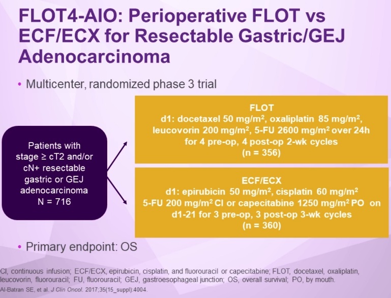
5-Fluorouracil 2600 mg/m² IV 24 h infusion day 1, Leucovorin (L) 200 mg/m² IV day 1, Oxaliplatin 85 mg/m² IV day 1, Docetaxel (T) 50 mg/m² IV day 1 (FLOT chemotherapy)


PRINCIPLES OF SYSTEMIC THERAPY

Systemic Therapy for Unresectable Locally Advanced, Recurrent or Metastatic Disease (where local therapy is not indicated)
• Trastuzumab should be added to first-line chemotherapy for HER2 overexpressing metastatic adenocarcinoma
--- Combination with fluoropyrimidine and cisplatin (category 1)
--- Combination with other chemotherapy agents (category 2B)
--- Trastuzumab is not recommended for use with anthracyclines

First-Line Therapy
• Two-drug cytotoxic regimens are preferred because of lower toxicity.
• Three-drug cytotoxic regimens should be reserved for medically fit patients with good PS and access to frequent toxicity evaluation.
Preferred Regimens
• Fluoropyrimidine (fluorouracil* or capecitabine) and cisplatin (category 1) • Fluoropyrimidine (fluorouracil* or capecitabine) and oxaliplatin
Other Recommended Regimens
• Paclitaxel with cisplatin or carboplatin
• Docetaxel with cisplatin
• Fluoropyrimidine (fluorouracil* or capecitabine)
• Docetaxel
• Paclitaxel
• Fluorouracil* ** and irinotecan
• DCF modifications
--- Docetaxel, cisplatin, and fluorouracil*
--- Docetaxel, oxaliplatin, and fluorouracil
--- Docetaxel, carboplatin, and fluorouracil (category 2B)
• ECF (epirubicin, cisplatin, and fluorouracil) (category 2B)
• ECF modifications (category 2B)
--- Epirubicin, oxaliplatin, and fluorouracil
--- Epirubicin, cisplatin, and capecitabine
--- Epirubicin, oxaliplatin, and capecitabine

* Leucovorin is indicated with certain fluorouracil-based regimens.
** Capecitabine cannot be used interchangeably with fluorouracil in regimens containing irinotecan.


Second-Line or Subsequent Therapy
• Dependent on prior therapy and PS

Preferred Regimens
• Ramucirumab and paclitaxel for adenocarcinoma (category 1 for EGJ adenocarcinoma; category 2A for esophageal adenocarcinoma)
• Docetaxel (category 1)
• Paclitaxel (category 1)
• Irinotecan (category 1)
• Fluorouracil* ** and irinotecan • Pembrolizumab --- For second-line or subsequent therapy for MSI-H or dMMR tumors

Other Recommended Regimens
• Ramucirumab for adenocarcinoma (category 1 for EGJ adenocarcinoma; category 2A for esophageal adenocarcinoma)
• Irinotecan and cisplatin
• Pembrolizumab
--- For third-line or subsequent therapy for PD-L1 positive esophageal and EGJ adenocarcinoma
• Docetaxel and irinotecan (category 2B)


Summary - Drug therapy of Esophageal Cancer

First Line for Advanced disease Her2 testing

Her2-negative:
        ◦ Chemotherapy

Her2-positive (Adenocarcinoma):
        ◦ Trastuzumab + Chemothrapy

Second Line MSI testing

MSS and mild neuropathy:
        ◦ Ramucirumab + Paclitaxel

MSI-H:
        ◦ Pembrolizumab

Third Line PDL1 testing

PDL1 negative: ◦ Chemotherapy
PDL1 positive:
        ◦ Pembrolizumab

Further somatic profiling.


Chemoradiation Regimens

◦ carboplatin + paclitaxel
◦ cisplatin + fluoropyrimidine
◦ cisplatin + gemcitabine (Squamous ca.)
◦ oxaliplatin + fluoropyrimidine (Adenocarcinoma)
◦ CROSS study: carboplatin + paclitaxel + Concurrent radiation

Chemotherapy Regimens
For Squamous cell carcinoma:
1. cisplatin + gemcitabine
2. carboplatin + paclitaxel
For Adenocarcinoma:
1. Oxalinplatin + 5FU
2. Ramucirumab + paclitaxel
3. Capecitabine + irinotecan


Treatment

Definitive Chemoradiation
For patients who are deemed either medically inoperable or have tumors that are unresectable, the efficacy of definitive chemoradiation has been established in numerous randomized controlled trials. For patients with squamous cell carcinomas of the esophagus, definitive chemoradiation may offer equivalent outcomes compared with preoperative chemoradiation followed by surgical resection.

Evidence (definitive chemoradiation):

A Radiation Therapy Oncology Group trial (RTOG-8501) randomly assigned patients to chemotherapy and radiation therapy versus radiation therapy alone. Patients were randomly assigned to receive radiation therapy alone (64 Gy in 32 fractions) or chemoradiation (50 Gy in 25 fractions) with concurrent cisplatin (75 mg/m2) and continuous-infusion 5-FU (1,000 mg/m2 on days 1 to 4 in weeks 1 and 5 followed by two additional cycles of chemotherapy administered 3 weeks apart).[Level of evidence: 1iiA]
There was an improvement in 5-year survival for the combined modality group (27% vs. 0%).
An 8-year follow-up of this trial demonstrated an OS rate of 22% for patients receiving chemoradiation therapy.

Intergroup-0123 (RTOG-9405 [NCT00002631]) was conducted in an attempt to improve upon the results of RTOG-8501. Intergroup-0123 randomly assigned 236 patients with localized esophageal tumors to undergo chemoradiation with high-dose radiation therapy (64.8 Gy) and four monthly cycles of 5-FU and cisplatin versus conventional-dose radiation therapy (50.4 Gy) and the same chemotherapy schedule.[Level of evidence: 1iiA]
Although originally designed to accrue 298 patients, this trial was closed in 1999 after a planned interim analysis showed that it was statistically unlikely that there would be any advantage to using high-dose radiation.
At a 2-year median follow-up, no statistically significant differences in median survival were observed between the high-dose and conventional-dose radiation therapy arms (13 months vs. 18 months), 2-year survival (31% vs. 40%), or local and regional failures (56% vs. 52%).
There was a higher treatment mortality in the higher-dose arm (9% vs. 2%); however, 7 of 11 deaths in the high-dose arm occurred in patients who had received 50.4 Gy or less.

An Eastern Cooperative Oncology Group trial (EST-1282) evaluated 135 patients.[Level of evidence: 1iiA]
This trial showed that chemotherapy plus radiation therapy provided a better 2-year survival rate than did radiation therapy alone, similar to results from the intergroup trial.

The PRODIGE5/ACCORD17 (NCT00861094) trial sought to evaluate and compare the efficacy and safety of oxaliplatin, fluorouracil, and leucovorin calcium (FOLFOX) versus 5-FU and cisplatin as the chemotherapy backbone among patients treated with definitive chemoradiation for localized esophageal cancer. In this multicenter, randomized, phase II and III trial, 267 patients were randomly assigned to receive either six cycles of FOLFOX (three cycles concomitant with radiation therapy), oxaliplatin (85 mg/m2), leucovorin (200 mg/m2), bolus 5-FU (400 mg/m2) and infusion 5-FU (1,600 mg/m2 over 46 hours) or four cycles of 5-FU (1,000 mg/m2 for 4 days) and cisplatin (75 mg/m2 on day 1). All patients received radiation therapy (50 Gy in 25 fractions).[Level of evidence: 1iiDiii]
With a median follow-up of 25.3 months, there was no significant difference in PFS (9.7 months with FOLFOX vs. 9.4 months with 5-FU and cisplatin; P = .64).
There was one death caused by toxicity in the FOLFOX group versus six deaths in the 5-FU and cisplatin arm (P = .066).
There were no significant differences in grade 3 or 4 adverse events between treatment groups. Among toxicities of all grades, paresthesia, sensory neuropathy, and increases in aspartate transaminase and alanine transaminase were more common in the FOLFOX group; whereas, increases in serum creatinine, mucositis, and alopecia were more common in the 5-FU and cisplatin group.
A phase III German trial also compared induction chemotherapy (three courses of bolus 5-FU, leucovorin, etoposide, and cisplatin) followed by chemoradiation therapy (cisplatin, etoposide, and 40 Gy) followed by surgery (arm A), or the same induction chemotherapy followed by chemoradiation therapy (at least 65 Gy) without surgery (arm B) for patients with T3 or T4 squamous cell carcinoma of the esophagus. OS was the primary outcome.[Level of evidence: 1iiA]
The analysis of 172 eligible, randomly assigned patients showed that OS at 2 years was not statistically significantly different between the two treatment groups (arm A, 39.9%; 95% CI, 29.4%–50.4%; arm B, 35.4%; 95% CI, 25.2%–45.6%; log-rank test for equivalence with 0.15, P < .007).
Local PFS was higher in the surgery group (2-year PFS, 64.3%; 95% CI, 52.1%–76.5%) than in the chemoradiation therapy group (2-year PFS, 40.7%; 95% CI, 28.9%–52.5%; HR for arm B vs. arm A, 2.1; 95% CI, 1.3–3.5; P < .003).
Treatment-related mortality was higher in the surgery group (12.8%) than in the chemoradiation therapy group (3.5%) (P < .03).

FFCD 9102 (NCT00416858) randomly assigned 259 patients with T3N0–1M0 thoracic esophageal cancer to receive either two cycles of 5-FU and cisplatin (days 1–5 and 22–26) and either conventional radiation therapy (46 Gy in 4.5 weeks) or split course (15 Gy, days 1–5 and 22–26). Patients with response were then randomly assigned to receive either surgical resection (arm A) or continuation of chemoradiation (arm B: three cycles of 5-FU plus cisplatin and either conventional 20 Gy or split-course 15 Gy radiation therapy).[Level of evidence: 1iiA]
Of the 259 randomly assigned patients, 230 (89%) had squamous cell carcinoma, and 29 patients (11%) had adenocarcinomas. The 2-year OS was 34% in patients randomly assigned to receive surgery versus 40% in patients randomly assigned to receive definitive chemoradiation (HR, 0.90; P = .44). Median survival was 17.7 months for surgery and 19.3 months for definitive chemoradiation.
The 3-month mortality rate was 9.3% in the surgery arm compared with 0.8% in the chemoradiation arm (P = .002).

Current Treatment of Esophageal Cancer and Promising Clinical Trials Underway

Siewert type I and type II tumors are to be esophageal tumors
With regard to adjuvant and neoadjuvant treatment of esophageal and gastroesophageal adenocarcinomas, we very much continue to administer preoperative chemoradiation. In general, treatment is given preoperatively. It is usually very difficult to give adjuvant treatment, but particularly so after an esophagectomy.
Esophageal adenocarcinoma: Preoperative chemoradiation + surgery. Complete response rates with chemoradiation alone are quite low, between 20% and 25%.
Esophageal squamous cell cancer: Definitive chemoradiation with observation. Pathologic response rate of almost 50%.

A potential alternative for adenocarcinoma tumors at the gastroesophageal junction is perioperative chemotherapy. Studies of perioperative chemotherapy for gastric cancer, such as the MAGIC and FLOT4 studies, have also included patients with gastroesophageal junction cancers. We still prefer to give chemoradiation to these individuals. This is mainly because, when the tumor is involving the esophagus and the gastroesophageal junction, a complete or R0 resection is achieved only about 70% of the time. The R0 resection rate is up to 90% with chemoradiation. However, in select patients, perioperative chemotherapy for gastroesophageal junction adenocarcinoma can also be considered, but such a decision should be made following a multidisciplinary review.

What therapy options are available for patients with advanced esophageal tumors?
Treatment differs for adenocarcinomas vs squamous cell carcinomas. With the exception of the KEYNOTE-181 study, treatment of squamous esophageal tumors has not really changed over the last 30 to 40 years and primarily consists of chemotherapy. The same chemotherapy drugs are active in both histologies: fluoropyrimidine, platinum agents, taxanes, and irinotecan. The normal sequence, throughout much of the world, is to consider a fluoropyrimidine/platinum doublet in the first-line setting; in the United States, the most widely used regimen is FOLFOX
In the second- or third-line setting, we would consider either a taxane or irinotecan.

Targeted therapies for adenocarcinomas
In 2010, based on the results of the Trastuzumab for Gastric Cancer (ToGA) study, trastuzumab was approved as the first targeted therapy for use in gastroesophageal and gastric cancer. Trastuzumab is used to treat tumors that are human epidermal growth factor receptor 2 (HER2)-positive (about 20% to 25% of tumors). The HER2 positivity rate is even higher in distal esophageal and gastroesophageal junction adenocarcinomas than it is in gastric cancer. So, trastuzumab in combination with FOLFOX is the standard of care for all HER2-positive esophageal or gastric adenocarcinomas.

in 2014, the anti-VEGF receptor antibody ramucirumab was approved in combination with paclitaxel in the second-line setting. Again, the approval was based on a gastric cancer study that also included patients with gastroesophageal junction adenocarcinomas. Since we now know that adenocarcinomas of the distal esophagus, gastroesophageal junction, and proximal stomach are molecularly the same based on the results of The Cancer Genome Atlas analysis,[5] we extrapolate that same data to distal esophageal adenocarcinomas as well.

In September 2017, the FDA approved pembrolizumab based on the results of the KEYNOTE-059 trial. This single-arm study evaluated almost 260 patients who received pembrolizumab as third-line or greater therapy. In patients with PD-L1–positive tumors, there did appear to be modest benefits; specifically, the response rate in this treatment-refractory patient population was about 12%. There was also durable survival: the 12-month survival rate was approximately 25% in a treatment-refractory population.
the FDA approval of pembrolizumab is contingent on the tumor cells being PD-L1–positive. This is determined using a specific assay with the 22C3 antibody. PD-L1 positivity is defined as a combined positive score of at least 1, which indicates that there is some staining in either the tumor cells or the peritumoral mononuclear cells.
(Note: On July 30, 2019, the Food and Drug Administration approved pembrolizumab (KEYTRUDA, Merck) for patients with recurrent, locally advanced or metastatic, squamous cell carcinoma of the esophagus (ESCC) whose tumors express PD-L1 (Combined Positive Score [CPS] ≥10), as determined by an FDA-approved test, with disease progression after one or more prior lines of systemic therapy.
FDA also approved a new use for the PD-L1 IHC 22C3 pharmDx kit as a companion diagnostic device for selecting patients for the above indication.)